Over the shoulder shot of pathologist using digital microscope at pathology department to detect congenital malformations

CASE STUDY

Pathology: Enhancing Diagnostic Accuracy with Digital Microscopes

Discover how Rigshospitalet's Pathology Department enhances diagnostic accuracy and efficiency using digital microscopes for detecting congenital malformations.

Overview

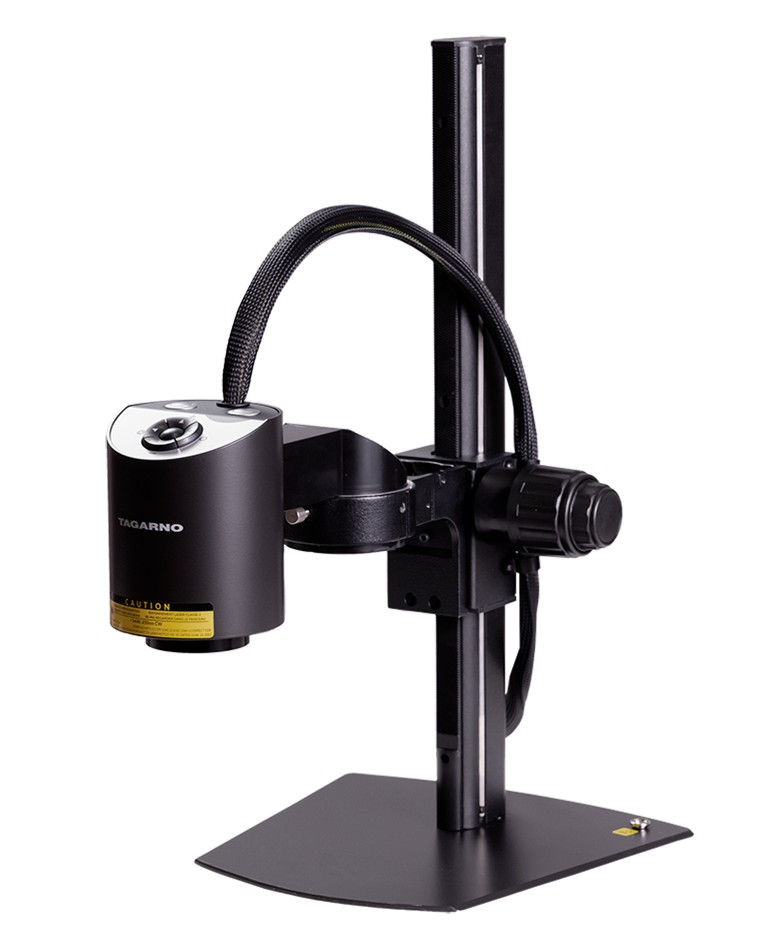
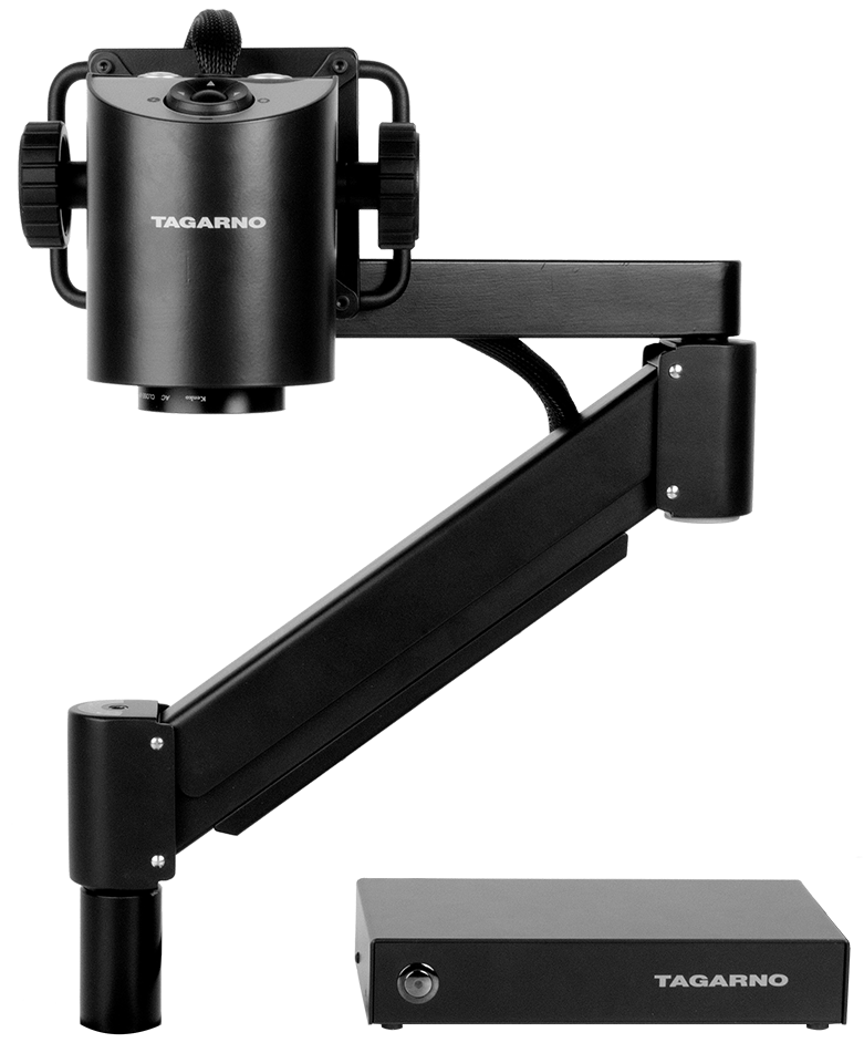
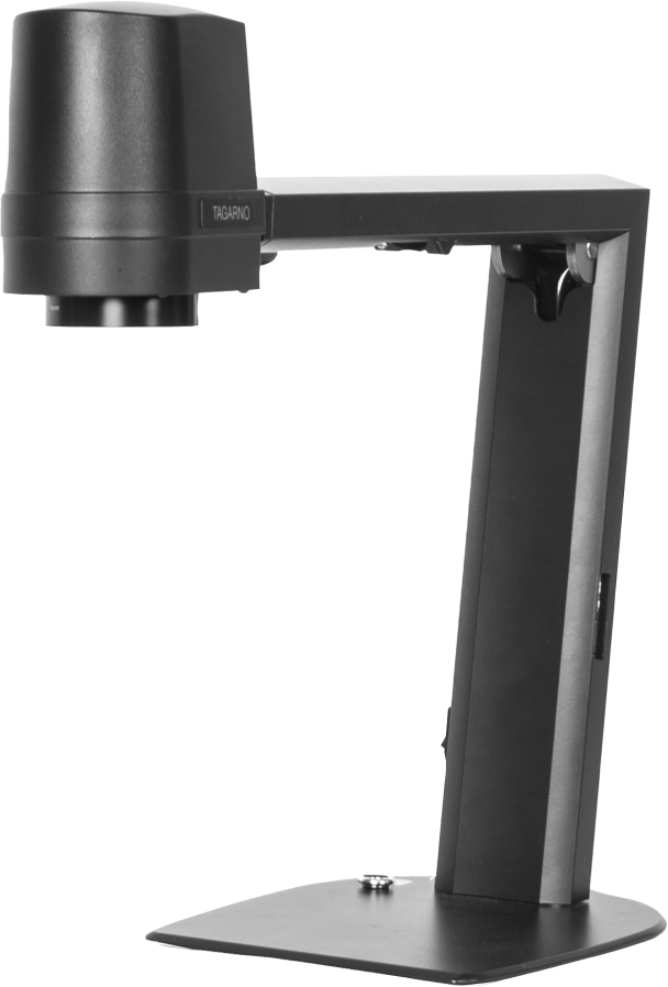
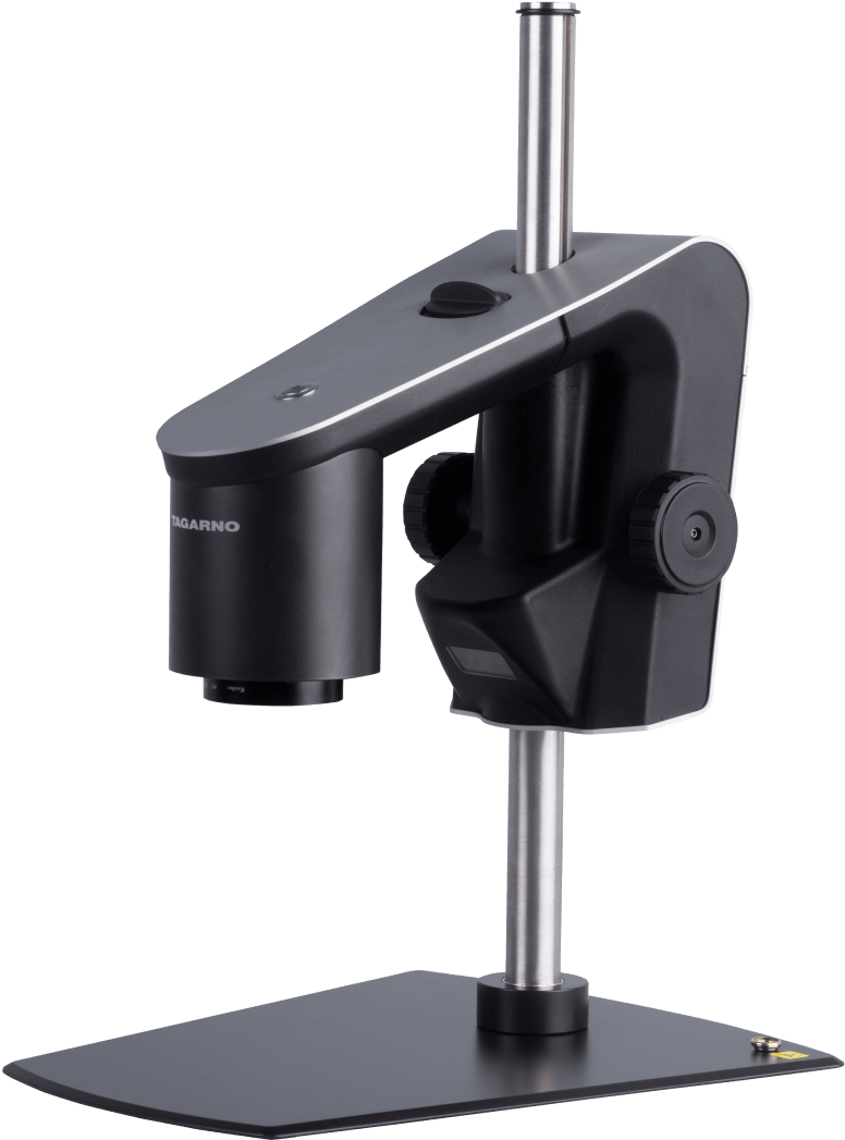
In 2023, the Pathology Department at Rigshospitalet invested in a digital microscope to enhance their diagnostic capabilities, particularly for autopsies. This investment was driven by the need for detailed examination of small organs and the importance of accurate image documentation to guide families through pregnancy loss.
Challenges

Autopsies require meticulous examination of small organs to determine causes of congenital malformations. Traditional methods posed significant challenges:

  • Detailed Examination: Accurate analysis of small organs was difficult without advanced imaging.
  • Collaboration: Effective supervision and collaboration with specialized doctors were limited.
  • Documentation: Capturing precise image documentation for individual cases was challenging.
  • Training: Training new colleagues effectively using traditional methods was inefficient.

Solution

The adoption of a digital microscope provided several benefits:

  • Enhanced Examination: The digital microscope allowed for better examination of small organs, improving diagnostic accuracy.
  • Remote Supervision and Collaboration: The ability to conduct supervision via Teams and collaborate with specialized doctors throughout the process enhanced the diagnostic workflow.
  • Image Documentation: The microscope enabled precise image capture for individual cases, crucial for guiding families through pregnancy loss.
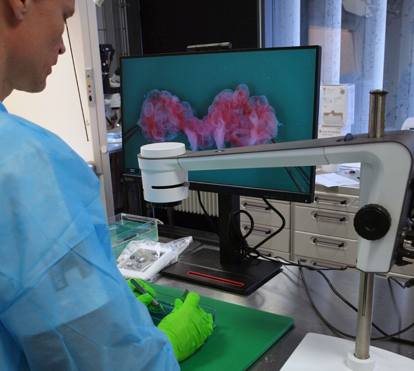
  • Training and Education: Live examinations and recorded videos for training new colleagues and creating educational material improved the learning experience.

Results

The Pathology Department at Rigshospitalet observed significant improvements:

  • Improved Diagnostic Accuracy: Enhanced examination capabilities led to more accurate diagnoses of congenital malformations.
  • Better Collaboration: Remote supervision and real-time collaboration with specialized doctors improved the diagnostic process.
  • Comprehensive Documentation: High-quality image documentation supported better case analysis and communication with families.
  • Effective Training: Utilizing live and recorded examinations for training purposes enriched the educational process for new colleagues.

Conclusion

The investment in a digital microscope has transformed the diagnostic capabilities at Rigshospitalet’s Pathology Department, particularly for detecting congenital malformations. This technology has not only improved diagnostic accuracy and collaboration but also enhanced documentation and training processes, ultimately providing better guidance for families through pregnancy loss.


You are now entering our German TAGARNO site Do you wish to continue?
No, stay here Yes, proceed